Nursing Management of a Deep Wound in the Sacral Site
Maculotti D and Dario Dassenno
Maculotti D1* and Dario Dassenno2
1Surgical Department, Ostomy Surgery Fondazione Poliambulanza, Brescia, Italy
2Catholic University of Sacro Cuore, Rome, Italy
- *Corresponding Author:
- Maculotti D Ambulatorio Stomizzati
Surgical Department, Ostomy Surgery Fondazione Poliambulanza, Brescia, Italy
Tel: (954)-214-3743
E-mail: danila.maculotti@poliambulanza
Received date: February 06, 2016, Accepted date: February 17, 2016; Published date: February 23, 2016
Citation: Maculotti D, Dassenno D. Nursing Management of a Deep Wound in the Sacral Site. Colorec Cancer 2016, 2:1. doi: 10.21767/2471-9943.100014
Abstract
Aim: Case report of a deep wound in the sacral site, (anterior-posterior perineal), caused by Cloacal neoplasia, with an urinary fistula. The objective was avoid critical colonizations and systemic infections after an abdominal resection and coccygectomy surgery.
Method: At first we used a iodoform gauze. In a second moment we treated the wound with a dressing of sodium carboxymethylcellulose impregnated with ionic silver enhanced by ethylenediaminetetra-acetic acid di sodium salt and benzethonium chloride.
Result: There were no exudate and bacterial colonization, and also no infection. The sacral stump has been covered by epitelium.
Conclusion: The silver Hydrofiber dressing together with the EDTA and benzethodium has been decisive. We can’t use negative pressure therapy. In 8 months of the treatment the wound has been reduced.
Keywords
Nursing care; Wound care; Urinary fistula; Granulation; Advanced medication; Vac terapy
Introduction
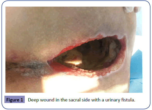
The clinical case report management of a deep sacral wound crater (anterio-posterior perineal), 26 cm length and 20 cm depth, in a woman experiencing a cloacla neoplasia. The patient was a young woman of 46 years old, she had an abdominoperineal resection and a coccygectomy surgery, hysteroannesectomy, permanent endcolostomy, urinary fistula (Figure 1).
Objectives
A urinary fistula became a complication in the wound management. The first objective of the treatment was to avoid crtitical colonizations and systemic infections.
Methods
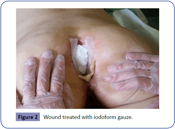
Step 1: In the first week the wound was packed on a daily base filling the cavity with a iodoform gauze. The wound appeared with an aboundant exudate inside the cavity with slaugh and necrotic tissue (Figure 2).
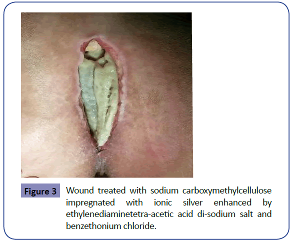
Step 2: After the wound was treated with a dressing of sodium carboxymethylcellulose impregnated with ionic silver enhanced by ethylenediaminetetra-acetic acid di-sodium salt and benzethonium chloride.
For 18 days the dressing has been changed on a daily base (Figure 3).
Results
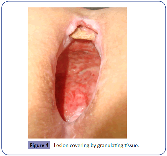
The dressing allowed the management of the exudates and bacterial colonization; clinical signs of localized or systemic infection has never been reported in the wound despite the presence of bacteria (Escherichia coli) and the urinary fistula [1-5]. In 8 months the wound reduced his dimension (8 cm length; 3 cm width; 6 cm depth); the wound bad has been maintained clean and the sacral stump was covered by granulating tissue (Figure 4).
Conclusion
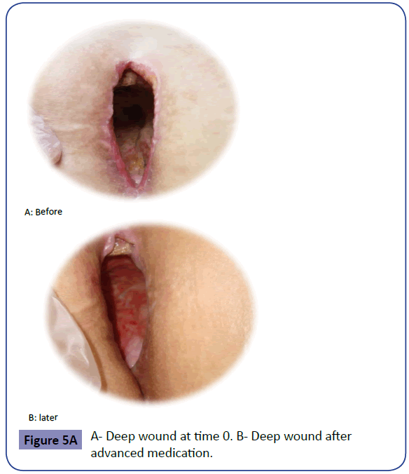
At first the urinary fistula didn’t allow the treatement with negative pressure therapy. The decision of treating the wound with advanced wound dressing has been a success. In 8 months the wound reduced his dimension without any infections of the wound bad with the evidence of an active tissue healing process (Figure 5).
References
- Walker M, Metcalf D, Parsons D, Bowler P (2015) A real-life clinical evaluation of a next-generation antimicrobial dressing on acute and chronic wounds. J Wound Care 24:11-22.
- Biffi R, Fattori L, Bertani E, Radice D, Rotmensz N, et al. (2012)Surgical site infections following colorectal cancer surgery: a randomized prospective trial comparing common and advanced antimicrobial dressing containing ionic silver. World JSurgOncol 10:94.
- Speck KE, Arnold MA, Ivancic V, Teitelbaum DH (2014) Cloaca and hydrocolpos: laparoscopic-, cystoscopic- and colposcopic-assisted vaginostomy tube placement. J PediatrSurg49:1867-1869.
- Greer SE, Duthie E, Cartolano B, Koehler KM, Maydick-Youngberg D, et al.(1999)Techniques for applying subatmospheric pressure dressing to wounds in difficult regions of anatomy. J Wound Ostomy ContinenceNurs 26: 250-253.
- Berdah SV, Mariette C, Denet C, Panis Y, Laurent C, et al.(2014) A multicentre, randomised, controlled trial to assess the safety, ease of use, and reliability of hyaluronic acid/carboxymethylcellulose powder adhesion barrier versus no barrier in colorectal laparoscopic surgery. Trial 15: 413.
Open Access Journals
- Aquaculture & Veterinary Science
- Chemistry & Chemical Sciences
- Clinical Sciences
- Engineering
- General Science
- Genetics & Molecular Biology
- Health Care & Nursing
- Immunology & Microbiology
- Materials Science
- Mathematics & Physics
- Medical Sciences
- Neurology & Psychiatry
- Oncology & Cancer Science
- Pharmaceutical Sciences
